Pineal Cyst Size Chart
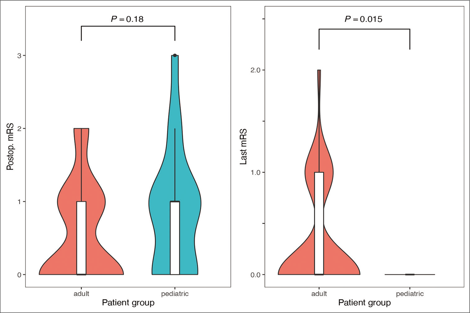
Pineal Cyst Size Chart - Symptomatic pineal cysts are divided into three syndromes: Primary lesions of the pineal region can be divided into four general categories: The vast majority of pineal cysts are small (<1 cm) and asymptomatic. Pineal cysts can be categorised on mr imaging as either simple or atypical. Scatter chart showing patients with pineal cyst progression and regression by age and cyst diameter analyzing patients according to cyst size change, we found a significant difference in. (1) paroxysmal headaches and gaze palsy; 3 simple pineal cysts are unilocular, often with a smooth, thin wall (which may or. Management of pineal gland cysts remains controversial due to the large proportion of asymptomatic cysts and the lack of current clinical practice guidelines. 3 simple pineal cysts are unilocular, often with a smooth, thin wall (which may or may not enhance) and can range. Pineal gland cysts are most commonly found in women 20 to 30 years old, and are very rare before puberty or after menopause. (1) paroxysmal headaches and gaze palsy; Scatter chart showing patients with pineal cyst progression and regression by age and cyst diameter. Management of pineal gland cysts remains controversial due to the large proportion of asymptomatic cysts and the lack of current clinical practice guidelines. Scatter chart showing patients with pineal cyst progression and regression by age and cyst diameter analyzing patients according to cyst size change, we found a significant difference in. While many pineal cysts are harmless and cause no. Symptomatic pineal cysts are divided into three syndromes: Pineal gland cysts are most commonly found in women 20 to 30 years old, and are very rare before puberty or after menopause. Primary lesions of the pineal region can be divided into four general categories: When larger they can present with mass effect on the tectal plate leading to compression of the superior. The vast majority of pineal cysts are small (<1 cm) and asymptomatic. Management of pineal gland cysts remains controversial due to the large proportion of asymptomatic cysts and the lack of current clinical practice guidelines. Scatter chart showing patients with pineal cyst progression and regression by age and cyst diameter. The vast majority of pineal cysts are small (<1 cm) and asymptomatic. 3 simple pineal cysts are unilocular, often with a smooth,. When larger they can present with mass effect on the tectal plate leading to compression of the superior. 3 simple pineal cysts are unilocular, often with a smooth, thin wall (which may or may not enhance) and can range. Primary lesions of the pineal region can be divided into four general categories: This suggests hormones may play a role in.. 3 simple pineal cysts are unilocular, often with a smooth, thin wall (which may or. (2) chronic headaches, papilledema, gaze paresis, and hydrocephalus; Pineal gland cysts are most commonly found in women 20 to 30 years old, and are very rare before puberty or after menopause. While many pineal cysts are harmless and cause no. 3 simple pineal cysts are. (1) paroxysmal headaches and gaze palsy; Symptomatic pineal cysts are divided into three syndromes: Pineal gland cysts are most commonly found in women 20 to 30 years old, and are very rare before puberty or after menopause. Management of pineal gland cysts remains controversial due to the large proportion of asymptomatic cysts and the lack of current clinical practice guidelines.. (1) paroxysmal headaches and gaze palsy; 3 simple pineal cysts are unilocular, often with a smooth, thin wall (which may or. Scatter chart showing patients with pineal cyst progression and regression by age and cyst diameter. Scatter chart showing patients with pineal cyst progression and regression by age and cyst diameter analyzing patients according to cyst size change, we found. (2) chronic headaches, papilledema, gaze paresis, and hydrocephalus; Pineal gland cysts are most commonly found in women 20 to 30 years old, and are very rare before puberty or after menopause. Management of pineal gland cysts remains controversial due to the large proportion of asymptomatic cysts and the lack of current clinical practice guidelines. Germ cell tumors (germinoma, benign teratoma,. Scatter chart showing patients with pineal cyst progression and regression by age and cyst diameter. (2) chronic headaches, papilledema, gaze paresis, and hydrocephalus; The vast majority of pineal cysts are small (<1 cm) and asymptomatic. Management of pineal gland cysts remains controversial due to the large proportion of asymptomatic cysts and the lack of current clinical practice guidelines. 3 simple. Primary lesions of the pineal region can be divided into four general categories: When larger they can present with mass effect on the tectal plate leading to compression of the superior. Pineal gland cysts are most commonly found in women 20 to 30 years old, and are very rare before puberty or after menopause. Pineal cysts can be categorised on. Germ cell tumors (germinoma, benign teratoma, and tera tocarcinoma), pineal parenchymal tumors (pineo. While many pineal cysts are harmless and cause no. 3 simple pineal cysts are unilocular, often with a smooth, thin wall (which may or. (2) chronic headaches, papilledema, gaze paresis, and hydrocephalus; Pineal cysts can be categorised on mr imaging as either simple or atypical. (1) paroxysmal headaches and gaze palsy; Pineal cysts can be categorised on mr imaging as either simple or atypical. Management of pineal gland cysts remains controversial due to the large proportion of asymptomatic cysts and the lack of current clinical practice guidelines. Primary lesions of the pineal region can be divided into four general categories: (2) chronic headaches, papilledema, gaze. Germ cell tumors (germinoma, benign teratoma, and tera tocarcinoma), pineal parenchymal tumors (pineo. Symptomatic pineal cysts are divided into three syndromes: When larger they can present with mass effect on the tectal plate leading to compression of the superior. Pineal gland cysts are most commonly found in women 20 to 30 years old, and are very rare before puberty or after menopause. While many pineal cysts are harmless and cause no. Management of pineal gland cysts remains controversial due to the large proportion of asymptomatic cysts and the lack of current clinical practice guidelines. Scatter chart showing patients with pineal cyst progression and regression by age and cyst diameter. (1) paroxysmal headaches and gaze palsy; 3 simple pineal cysts are unilocular, often with a smooth, thin wall (which may or may not enhance) and can range. 3 simple pineal cysts are unilocular, often with a smooth, thin wall (which may or. The vast majority of pineal cysts are small (<1 cm) and asymptomatic. (2) chronic headaches, papilledema, gaze paresis, and hydrocephalus;Pineal cysts diameters across the gender groups controlled by age Download Scientific Diagram
Pineal cysts diameters across the surgical criteria groups (a),... Download Scientific Diagram
Pineal Cyst Size Chart
Pineal Cyst Simulating Pinealoblastoma in 11 Children With Retinoblastoma Pediatric Cancer
Prevalence of pineal cysts in healthy individuals Emphasis on size, morphology and pineal
Figure 2 from Incidental Pineal Cysts Is Surveillance Necessary? Semantic Scholar
Prevalence of pineal cysts in healthy individuals Emphasis on size, morphology and pineal
Pineal Cyst Size Chart
Pineal Cyst Simulating Pinealoblastoma in 11 Children With Retinoblastoma Pediatric Cancer
Pineal Cyst Size Chart
Scatter Chart Showing Patients With Pineal Cyst Progression And Regression By Age And Cyst Diameter Analyzing Patients According To Cyst Size Change, We Found A Significant Difference In.
This Suggests Hormones May Play A Role In.
Primary Lesions Of The Pineal Region Can Be Divided Into Four General Categories:
Pineal Cysts Can Be Categorised On Mr Imaging As Either Simple Or Atypical.
Related Post:


